

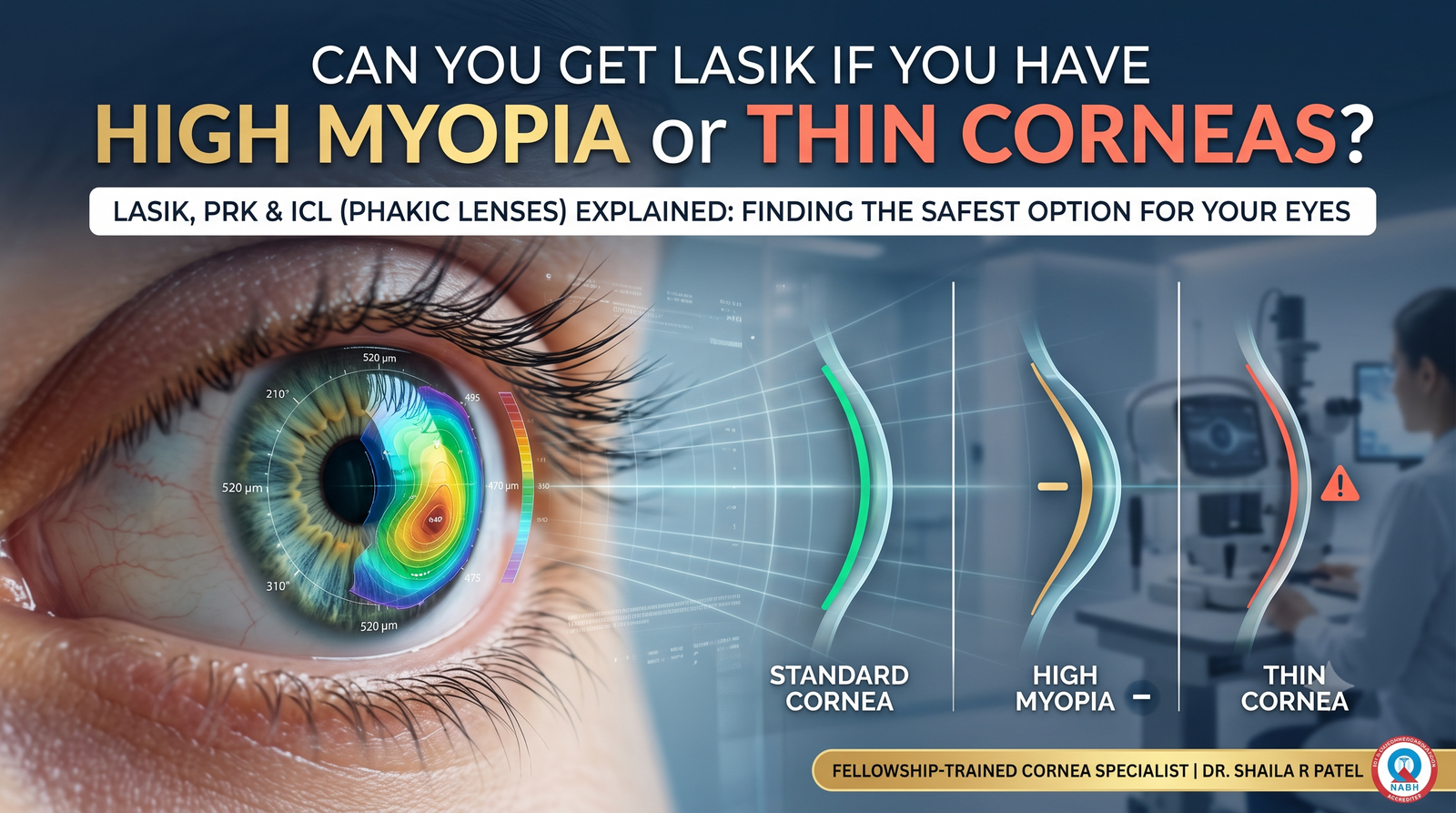
For millions of people living with severe nearsightedness, waking up without immediately reaching for thick glasses or wrestling with dehydrated contact lenses feels like an unattainable dream. When they finally walk into an eye clinic for a consultation, many are met with disheartening words: “Your corneas are too thin,” or “Your prescription is simply too high for LASIK.”
Hearing that you are not a candidate for traditional laser vision correction can feel like a closed door. However, in modern refractive surgery, being disqualified from LASIK does not mean you are disqualified from visual freedom.
Advances in ophthalmic engineering have made corneal thickness constraints largely irrelevant for a wide range of patients. The primary solution is the Phakic Intraocular Lens, most notably the EVO ICL (Implantable Collamer Lens).
Whether your spectacle power is $-8.00\text{ D}$, $-14.00\text{ D}$, or higher, or your corneal thickness falls well below the standard safety limits for laser ablation, phakic lens technology provides an alternative that preserves corneal architecture entirely.
This guide explains the biomechanics of why LASIK fails in thin or highly myopic eyes, how the ICL procedure works, its safety profiles, eligibility criteria, and how to determine whether this additive procedure is right for you.
1. The Physics of LASIK: Why Corneal Tissue Limits the Laser
To understand why a surgeon might advise against LASIK, you must first understand how an excimer laser alters the eye.
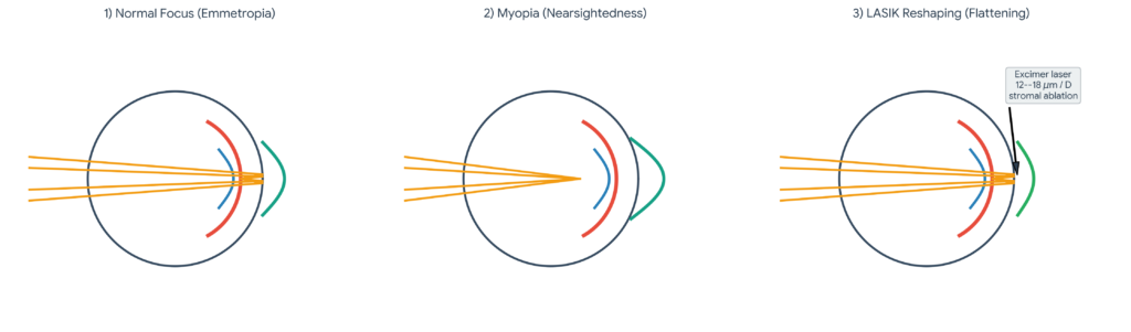
The cornea is the eye’s clear, dome-shaped front window, averaging 530 to 550 microns ($\mu\text{m}$) in central thickness—roughly the thickness of five sheets of paper. It provides around 70% of the eye’s total optical focusing power.
In a nearsighted eye, the eyeball is either too long from front to back, or the cornea curves too steeply. In both instances, incoming light rays come to a sharp focus in front of the retina rather than directly on its sensory surface, creating distant blur.
The Math Behind Laser Tissue Ablation
LASIK corrects nearsightedness by vaporizing microscopic amounts of tissue from the middle layer of the cornea (the stroma) using a 193 nm ultraviolet excimer laser. This flattens the central corneal curvature, pushing the focal point back onto the retina.
- To correct $1.00\text{ Diopter (D)}$ of myopia, the laser must permanently remove approximately 12 to 18 microns ($\mu\text{m}$) of corneal stroma (governed by the Munnerlyn formula and optical ablation zone diameter).
- If your prescription is $-4.00\text{ D}$, the laser removes roughly $60\text{ to }70\ \mu\text{m}$ of tissue.
- If your prescription is $-10.00\text{ D}$, the laser would need to consume $140\text{ to }180\ \mu\text{m}$ of stromal tissue.
The Structural Safety Thresholds (RSB and PTA)
A responsible refractive surgeon will never compromise the structural stability of the cornea. Two parameters guide surgical decisions:
- The Flap Consumption: In Femto-LASIK, creating the surface flap consumes roughly $90\text{ to }110\ \mu\text{m}$ of anterior tissue. Because this flap is cut, it no longer contributes meaningfully to the biomechanical tensile strength of the corneal wall.
- Residual Stromal Bed (RSB): The unablated, untouched bed of stroma beneath the flap must remain thick enough to withstand the eye’s internal fluid pressure (Intraocular Pressure or IOP). Worldwide safety standards mandate an absolute minimum RSB of $250\ \mu\text{m}$, with most conservative corneal specialists demanding $280\text{ to }300\ \mu\text{m}$.
- Percent Tissue Altered (PTA):

If the PTA value exceeds 40%, the mathematical risk of long-term structural failure escalates significantly.
| Parameter | Value / Calculation | Status & Clinical Assessment |
|---|---|---|
| Starting Central Corneal Thickness | 510 µm | Slightly thin cornea |
| Minus Femto-LASIK Flap Thickness | – 100 µm | Standard flap creation |
| Minus Tissue Ablation for -9.00 D | – 135 µm | Based on an average of ~15 µm per diopter |
| Resulting Residual Stromal Bed (RSB) | 275 µm | CRITICAL RED ZONE! (Risk of ectasia) |
| Percent Tissue Altered (PTA) | (100 + 135) / 510 = 46.1% | High Risk (Threshold limit is generally < 40%) |
The Real Danger: Iatrogenic Corneal Ectasia
What happens if a surgeon ignores these safety limits and performs LASIK on a cornea that is naturally too thin or structurally over-ablated?
The remaining bed of stromal tissue becomes too weak to withstand normal intraocular pressure. Over the following months or years, the cornea begins to stretch, thin, and bulge outward irregularly—a serious, vision-threatening condition known as iatrogenic post-LASIK corneal ectasia. Ectasia causes severe irregular astigmatism, distorted ghosting, and can ultimately require corneal cross-linking or full corneal transplantation.
This is why experienced cornea and refractive surgeons like Dr. Shaila Rajkumar Patel at Infinite Vision Care uphold strict screening criteria. If your corneas are under $490\ \mu\text{m}$, or your prescription exceeds the safe ablation limit, you are advised against LASIK to protect your long-term ocular health.
2. What Is an ICL (Implantable Collamer Lens)?
When laser surgery cannot safely reshape the cornea from the outside, the natural solution is to add a lens on the inside. This is the role of the Phakic Intraocular Lens (pIOL), most commonly known as the EVO ICL (STAAR Surgical).
“Phakic” vs. “Pseudophakic”: An Important Distinction
- Pseudophakic Lens (Cataract Surgery): The eye’s natural, aged internal crystalline lens is removed via ultrasound and replaced with an artificial intraocular lens (IOL).
- Phakic Lens (ICL): The word “phakic” means you still retain your natural crystalline lens. Nothing inside the eye is removed. Your natural lens remains untouched, allowing your eyes to retain their youthful dynamic focusing ability (accommodation) to see near, intermediate, and distant objects seamlessly.
What Is Collamer?
The EVO ICL is not made of plastic, glass, or ordinary silicone. It is manufactured from Collamer—a biocompatible material composed of 60% polyhydroxyethylmethacrylate (poly-HEMA), 36% water, 3.8% benzophenone (which provides natural UV protection), and 0.2% purified porcine collagen.
- Biocompatible Integration: Because Collamer contains purified collagen, the human body’s immune system does not recognize it as a foreign object. It does not cause rejection, chronic inflammation, or tissue scarring.
- Optical Transparency: Collamer has a high water content and a refractive index ($n = 1.45$) close to that of the natural human crystalline lens, eliminating internal reflections, scatter, and chromatic aberrations.
- Hydrophilic Cushion: The surface of Collamer carries a natural negative charge that attracts fibronectin molecules from the aqueous humor. This forms a protective protein monolayer across the lens, preventing cell adhesion and keeping the optical zone clear indefinitely.
3. The EVO ICL Revolution: Why Modern ICLs No Longer Require Laser Holes
Earlier generations of phakic lenses required an extra pre-operative step called a Peripheral Iridotomy (PI). A surgeon had to use an Nd:YAG laser to punch one or two tiny safety holes through the outer edge of your iris a week before surgery. This was necessary to ensure that fluid (aqueous humor) could circulate between the back of the eye and the front of the eye without becoming trapped and causing an acute spike in eye pressure (pupillary block glaucoma).
| FeatureOlder ICL GenerationsModern EVO ICL (CentraFLOW)Pre-Operative RequirementsRequires a painful pre-op YAG iridotomy to punch holes through the iris.No iridotomy required, eliminating the extra surgical step.Fluid Management MechanismRelies on surgical holes in the iris to prevent fluid buildup and glaucoma.Features a 360 µm central micro-aperture (KS-AquaPORT) for natural fluid flow.Associated RisksHigher risk of glare, halos, transillumination defects, and bleeding.Dramatically lowers the risk of optical side effects and structural trauma. |
|---|
The CentraFLOW Technology (KS-AquaPORT)
Modern surgeons use the EVO and EVO+ ICL family, which incorporates CentraFLOW technology.
In the exact center of the EVO ICL is a laser-engineered micro-hole measuring $360\ \mu\text{m}$ (0.36 mm) across—the KS-AquaPORT.
- This central port permits natural physiological circulation of aqueous humor directly through the center of the lens.
- It equalizes fluid pressure between the posterior and anterior chambers without altering the iris.
- Studies have confirmed that this micro-hole does not cause visual disturbances, glare, or diffraction rings, while safely bathing the front surface of your natural lens in nutrient-rich fluid.
4. Head-to-Head: LASIK vs. PRK vs. SMILE vs. ICL
When patients discover they have thin corneas or high myopia, they often ask about surface treatments like PRK or keyhole options like SMILE before considering an implantable lens.
| Clinical Metric | Femto-LASIK | PRK / TransPRK | SMILE / SILK | EVO ICL (Phakic Lens) |
| Surgical Method | Flap cut; stromal tissue vaporized | Surface scraped; stromal tissue vaporized | Keyhole cut; internal lenticule extracted | Tissue-additive; collamer lens implanted |
| Corneal Tissue Consumed | High ($\approx 15\ \mu\text{m}/\text{D} + 100\ \mu\text{m}\text{ flap}$) | Moderate ($\approx 15\ \mu\text{m}/\text{D}$; zero flap) | Moderate ($\approx 14\ \mu\text{m}/\text{D}$; zero flap) | Zero (0 µm corneal tissue removed) |
| Treatment Range (Myopia) | Up to $-8.00\text{ D}$ | Up to $-6.00\text{ D}$ | Up to $-10.00\text{ D}$ | $-0.50\text{ D}$ up to $-20.00\text{ D}$ |
| Treatment Range (Cylinder) | Up to $5.00\text{ D}$ | Up to $3.00\text{ D}$ | Up to $5.00\text{ D}$ | Up to $6.00\text{ D}$ (Toric ICL) |
| Minimum Cornea Required | $\ge 500\ \mu\text{m}$ | $\ge 460\text{–}480\ \mu\text{m}$ | $\ge 480\text{–}490\ \mu\text{m}$ | Any thickness (Cornea is irrelevant) |
| Dry Eye Induction | Moderate to High (3–6 months) | Low to Moderate | Low | Zero (Corneal nerves left intact) |
| Reversibility | Irreversible | Irreversible | Irreversible | 100% Completely Reversible |
| Recovery Speed | 24 Hours | 5 to 7 Days (Painful) | 24 to 48 Hours | 24 Hours (Immediate clarity) |
| Visual Quality in High Power | Prone to spherical aberration | Prone to haze/regression | Prone to minor contrast drop | High-Definition (Zero spherical shift) |
5. Why High Myopes Often Achieve Better Vision Quality with ICL than LASIK
A common misconception is that ICL is merely a “backup plan” for patients who fail LASIK screening. In reality, for patients with prescriptions above -6.00 or -7.00 Diopters ICL often provides superior optical clarity compared to laser ablation.
| Optical AttributeHigh-Power LASIK (Significant Flattening)High-Power ICL (Original Curvature Preserved)Corneal ShapeCarved severely flat, structurally altering the outer layer.Maintains its natural spherical curve completely untouched.Light RefractionLight rays bend awkwardly at the laser-treated peripheral edges.Light focuses uniformly through a pristine internal optic.Visual AberrationsInduces high spherical aberration, leading to night glare and halos.Zero induced corneal aberration, preserving sharp HD contrast and excellent night vision. |
|---|
The Problem with Flat Corneas
When an excimer laser treats a high prescription (such as -9.00, it must drastically flatten the center of the cornea. This transforms the natural prolate (steep center, flatter edge) shape of the human cornea into an unnatural oblate (flat center, steep edge) surface.
This steep transition zone induces spherical aberrations:
- Incoming light rays at the periphery of your pupil focus at a different point than rays entering through the center.
- The patient may achieve 20/20 vision on a black-and-white chart in a lit exam room, but in low-light conditions—such as driving at night—they experience halos, starbursts around headlights, and reduced contrast sensitivity.
- Image magnification drops: High spectacle and laser corrections shrink image size on the retina, whereas an intraocular lens positioned close to the nodal point of the eye preserves natural retinal image size.
The ICL Optical Advantage
Because an ICL does not alter corneal curvature, the natural prolate architecture of your eye is preserved. The lens optics are manufactured in a precision laboratory under strict cleanroom conditions, delivering sharp contrast, deep color saturation, and clear night vision without spherical aberration.
6. Are You a Candidate for ICL? (The Vital Safety Checklist)
Because an ICL is placed inside the eye rather than on its surface, candidacy depends on internal anatomical dimensions rather than corneal thickness.
During your comprehensive pre-operative evaluation at an advanced clinic like Infinite Vision Care, your surgeon will measure several parameters:
1. Anterior Chamber Depth (ACD)
The anterior chamber is the fluid-filled space between the back surface of your cornea (endothelium) and the front surface of your crystalline lens.
- The FDA and global guidelines require an internal ACD of at least $2.8\text{ to }3.0\text{ mm}$.
- If your eye is anatomically shallow ($< 2.8\text{ mm}$), there is insufficient clearance to safely house the lens without crowding internal structures. Fortunately, myopic eyes are naturally longer and deeper than average, so the vast majority of nearsighted patients easily meet this requirement.
2. Endothelial Cell Density (ECD)
The corneal endothelium is a non-regenerative monolayer of hexagonal cells lining the inner surface of the cornea. These cells pump fluid out of the cornea to maintain its clarity.
- Humans are born with roughly $4,000\text{ cells/mm}^2$, which naturally decline at a rate of about 0.5% per year.
- To safely qualify for ICL surgery, your baseline ECD must comfortably exceed age-stratified minimums (typically $\ge 2,000\text{ to }2,400\text{ cells/mm}^2$).
- Specular microscopy confirms your endothelial cell health before surgery.
3. Open Drainage Angles (Gonioscopy)
The drainage angle (trabecular meshwork) where the iris meets the cornea must be wide open—classified as Shaffer Grade III or IV. This ensures that fluid outflow remains unobstructed once the lens is positioned.
4. White-to-White (WTW) & Sulcus-to-Sulcus Sizing
The surgeon must measure the exact diameter of your eye—both externally (White-to-White or corneal diameter) and internally (Sulcus-to-Sulcus using high-resolution Anterior Segment OCT or Very High-Frequency Ultrasound Biomicroscopy / UBM).
This measurement determines the physical size of your custom ICL ($12.1\text{ mm}$, $12.6\text{ mm}$, $13.2\text{ mm}$, or $13.7\text{ mm}$). Proper sizing ensures the lens arches over your natural crystalline lens with an optimal vault.
7. What Happens on Surgery Day: The 15-Minute Procedure
The thought of an intraocular procedure can make patients nervous. In practice, the EVO ICL procedure is quick, gentle, and performed on an outpatient basis under topical drop anesthesia.
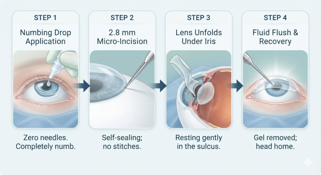
Step 1: Anesthetic Preparation (No General Anesthesia)
You will receive dilating eye drops and mild oral medication to help you relax. When you enter the sterile surgical theater, topical numbing eye drops are applied. Your eye is fully numbed within seconds; there are no retrobulbar injections, needles, or general anesthesia required.
Step 2: The Self-Sealing Micro-Incision
Under a high-definition surgical operating microscope, the surgeon makes a micro-incision measuring just $2.8\text{ to }3.0\text{ mm}$ at the clear peripheral border of the cornea. Because this incision is small and engineered with a stepped architecture, it self-seals through intraocular pressure without requiring stitches. A protective viscoelastic gel is injected to maintain chamber space.
Step 3: Lens Injection & Positioning
The foldable EVO ICL is loaded into a micro-cartridge. The surgeon gently injects the lens through the micro-incision. As it enters the anterior chamber, the Collamer lens unfurls over your pupil. Using a soft, blunt micro-manipulator, the surgeon tucks the four flexible footplates (haptics) of the lens behind your colored iris into the ciliary sulcus. The lens rests securely in place, invisible to the naked eye.
Step 4: Viscoelastic Washout
The surgeon washes out the viscoelastic gel using a balanced salt solution, checks your intraocular pressure, and instills antibiotic-steroid drops. The entire procedure takes 10 to 15 minutes per eye.
Both eyes are often treated on the same day or separated by 24 to 48 hours. After a short observation period to ensure stable eye pressure, you head home wearing protective eyewear.
8. Recovery Timeline and Life After ICL
One of the most satisfying aspects of ICL surgery is the immediate visual recovery. Because no corneal flap was cut and no surface tissue was scraped away, healing is rapid and largely pain-free.
- Hours 1 to 4: As the numbing drops wear off, your eyes may feel watery, heavy, or mildly gritty. Resting in a dimly lit room with your eyes closed is recommended.
- Day 1 (The Next Morning): Most patients wake up with 20/20 or 20/15 functional vision. You will visit the clinic for your first post-op check, where the surgeon measures your eye pressure and assesses the lens “vault” under a slit-lamp microscope. You can read, use your phone, and resume light desk work.
- Week 1: You will use prescribed antibiotic and anti-inflammatory eye drops to support healing. Light gym workouts, walking, and office routines can resume. Avoid getting tap water directly in your eyes or rubbing them.
- Week 4: The micro-incisions are completely healed. All physical restrictions—including swimming, weightlifting, intense sports, and eye makeup—are lifted.
9. Understanding the “Vault” and Long-Term Safety
When researching ICL online, you may encounter clinical discussions about the vault.
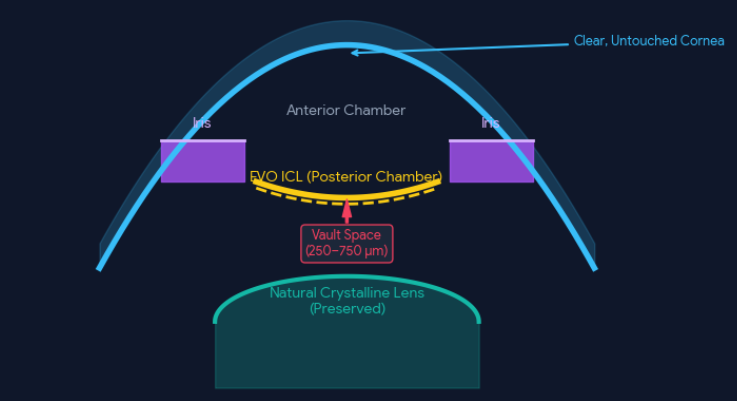
The vault is the vertical clearance gap between the posterior (back) surface of the ICL and the anterior (front) surface of your eye’s natural crystalline lens. Maintaining an appropriate vault is key to long-term safety:
The Ideal Vault Window: $250\ \mu\text{m}\text{ to }750\ \mu\text{m}$
- If the Vault is Too Low ($< 150\text{–}250\ \mu\text{m}$): If the ICL sits too close to or touches the natural crystalline lens, it can disrupt nutrient exchange in the anterior lens capsule, increasing the risk of anterior subcapsular cataract formation.
- If the Vault is Too High ($> 750\text{–}1000\ \mu\text{m}$): If the ICL arches too far forward, it can push the iris forward, narrowing the drainage angle and increasing the risk of elevated eye pressure (glaucoma) or endothelial cell contact.
Why the EVO Design Transformed Vault Safety
In older generations of ICLs, anterior subcapsular cataracts occurred in 1% to 2% of patients due to low vaults and stagnant aqueous flow.
With the introduction of the EVO ICL’s central KS-AquaPORT, aqueous fluid circulates continuously across the front of the natural lens. In FDA clinical trials of the EVO ICL, zero eyes developed anterior subcapsular cataracts during the observation period.
Furthermore, high-frequency ultrasound and optical coherence tomography (OCT) allow modern surgeons to size the lens accurately before surgery, keeping the vault within the safe target window.
10. The Ultimate Safety Feature: 100% Reversibility
LASIK, PRK, SMILE, and Contoura Vision are permanent, subtractive procedures. Once corneal stroma is vaporized or an internal lenticule is extracted, that tissue is gone forever. If you are unhappy with the optical outcome, the procedure cannot be undone.
| Feature | Laser Vision Correction (LASIK / PRK / SMILE / CONTOURA) | EVO ICL Surgery |
|---|---|---|
| Surgical Approach | SUBTRACTIVE | ADDITIVE |
| Tissue Impact | Corneal tissue is permanently removed by an excimer or femtosecond laser to change refraction. | Zero tissue is removed from the cornea or any other internal eye structure. |
| Reversibility | Irreversible change to the structural architecture and biomechanics of the eye. | Completely reversible; the lens can be safely removed or upgraded at any time if your prescription changes. |
The ICL is an additive, reversible technology.
- If your prescription shifts substantially in the future, the lens can be removed and exchanged.
- If you develop cataracts in your 60s or 70s, your surgeon can remove the ICL through a micro-incision in two minutes, leaving your eye in its natural state to proceed with standard cataract surgery and premium IOL implantation.
- If advanced refractive technology becomes available down the road, your natural optical pathways remain unmodified.
For patients who feel hesitant about permanently altering their corneas, this reversibility provides reassurance.
11. What Does ICL Cost Compared to LASIK?
Cost is a frequent consideration when evaluating refractive options. Globally and across India, ICL surgery is more expensive than standard LASIK or Contoura Vision. Understanding the factors behind this difference helps explain why.
Cost Breakdown Table (India & Global Averages)
| Procedure | Cost Range in India (Both Eyes) | Why the Price Differs |
| Standard Femto-LASIK | ₹50,000 – ₹85,000 | Uses excimer & femtosecond laser platform licenses. |
| Contoura Vision (Topography) | ₹75,000 – ₹1,15,000 | Advanced topography mapping & license royalties. |
| SMILE Pro / SILK | ₹90,000 – ₹1,60,000 | Single-use patient interface packs & laser technology. |
| EVO ICL (Spherical) | ₹1,20,000 – ₹1,60,000 | Custom Swiss-manufactured Collamer implants. |
| EVO Toric ICL (Astigmatism) | ₹1,50,000 – ₹2,00,000 | Custom-manufactured astigmatic optics. |
Why Does ICL Carry a Premium Price?
- Custom Swiss Fabrication: Each EVO ICL is manufactured by STAAR Surgical in Switzerland to match your individual refraction, corneal diameter, and anterior chamber dimensions.
- Toric Customization: If you have astigmatism alongside myopia, your Toric ICL is custom-milled with precise rotational axis parameters.
- Surgical Consumables: The procedure uses specialized ophthalmic viscoelastic devices, intraocular instruments, and sterile injector cartridges in a certified surgical theater.
- Microsurgical Expertise: Implanting an intraocular lens requires the skill of a fellowship-trained anterior segment or corneal surgeon.
When viewed as an investment in lifelong visual freedom without dry eye complications or tissue destruction, many patients with high prescriptions find the visual quality and peace of mind well worth the initial cost.
12. Frequently Asked Questions (FAQs)
Q1: Can an ICL be seen by other people?
No. Because the EVO ICL sits behind your colored iris and pupil, it is invisible to the naked eye. Even friends standing close to you will not be able to see it. Only an eye care professional looking through a high-magnification slit-lamp microscope can detect it.
Q2: Can I feel the lens inside my eye?
No. The cornea surface is rich with sensory nerve endings, which is why a speck of dust or a contact lens causes discomfort. The interior posterior chamber where the ICL rests has no comparable sensory nerve receptors. Once the lens is placed, you will not feel it when blinking, moving your eyes, or sleeping.
Q3: Can an ICL slip out of place or fall into the back of my eye?
No. The ICL is positioned in the ciliary sulcus behind the iris. The anatomical structures of the iris and zonular fibers create a secure natural pocket that prevents the lens from moving forward or falling backward.
Q4: Will I still get presbyopia (reading glasses) when I turn 40?
Yes. Presbyopia is the natural, age-related stiffening of the eye’s internal crystalline lens, making it harder to focus on close objects over time. Because the ICL preserves your natural lens, it does not stop this process. Around age 45, you may still need reading glasses for close-up print, or your surgeon can adjust your optical strategy accordingly.
Q5: Is ICL approved by health insurance?
In many regions, including India, health insurance policies consider refractive surgery elective. However, if your prescription exceeds high-myopia thresholds (often -7.50 or higher), some private insurers cover ICL implantation under medical necessity clauses. It is advisable to consult your provider’s claims desk during your evaluation.
The Path Forward: Choosing Expert Care
Being told your corneas are too thin for LASIK can initially feel disappointing, but it is often a blessing in disguise. It keeps you from undergoing an over-aggressive laser ablation that could compromise your corneal stability, directing you toward a safer, tissue-preserving option.
At Infinite Vision Care and Laser Centre, Dr. Shaila Rajkumar Patel brings fellowship-trained expertise in Cornea, Cataract, and Refractive Surgery from the world-renowned Aravind Eye Hospital. Having authored chapters in premier international references like the Yanoff Textbook of Ophthalmology, Dr. Patel combines academic rigor with patient-first care:
- Comprehensive Safety Screening: In-house corneal tomography, anterior chamber depth biometry, and endothelial specular microscopy.
- NABH-Accredited Excellence: Modern surgical suites equipped for both laser vision correction and phakic IOL implantation.
- Objective Clinical Guidance: If your eyes suit Contoura Vision or SILK, you will receive clear guidance; if your corneas require the protection of an EVO ICL, you will be paired with the safest solution for your eyes.
You do not have to settle for heavy glasses or dry contact lenses. Discover how modern phakic lens technology can transform your eyesight. Schedule your consultation with a fellowship-trained Ophthalmologist in Mira Road and Ophthalmologist in Kandivali at Infinite Vision Care today.Being told your corneas are too thin for LASIK can initially feel disappointing, but it is often a blessing in disguise. It keeps you from undergoing an over-aggressive laser ablation that could compromise your corneal stability, directing you toward a safer, tissue-preserving option
